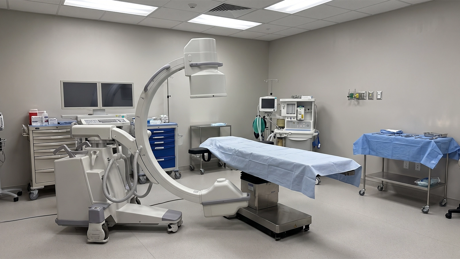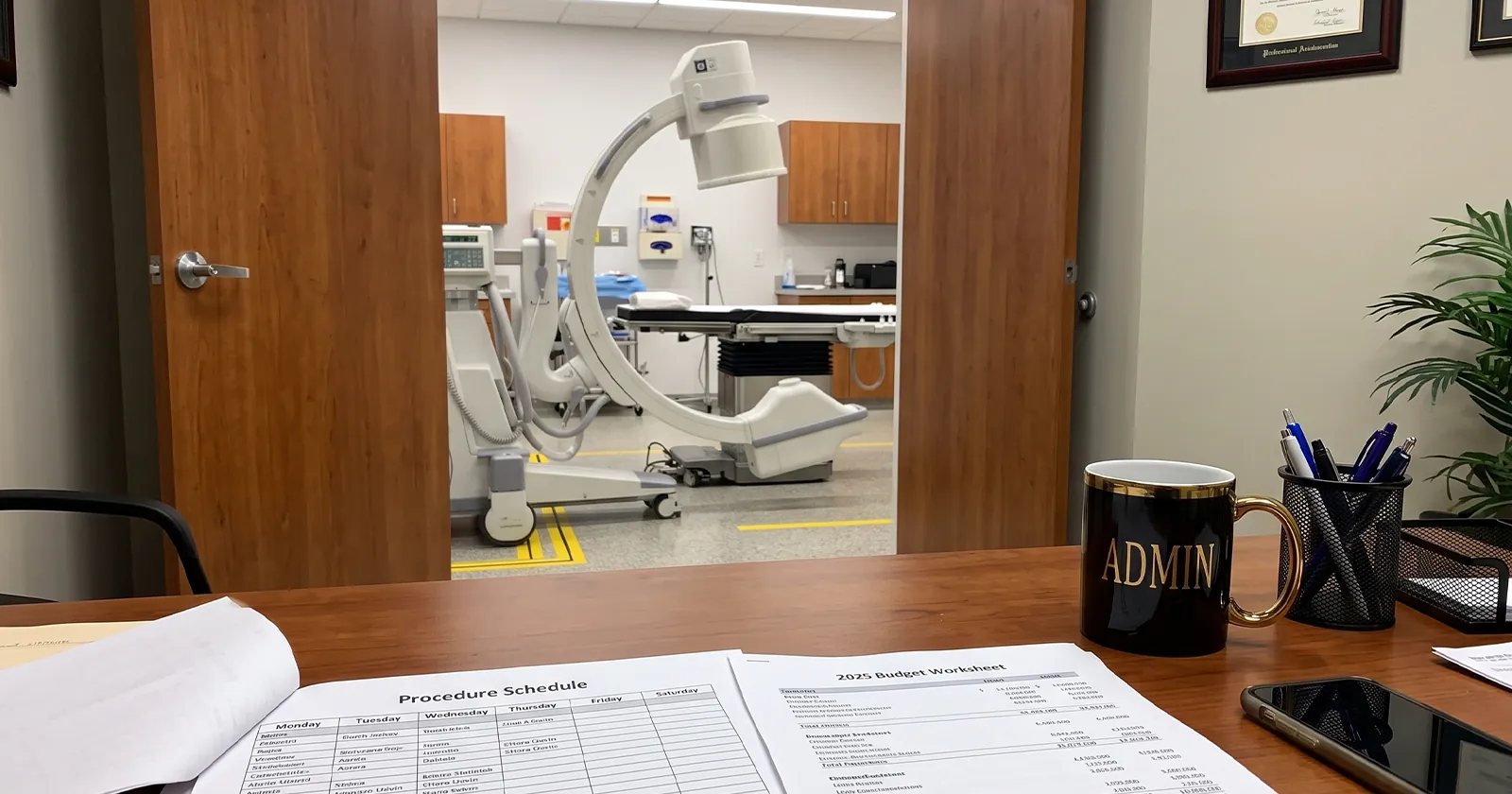
Anyone who’s stood in an OR while a C-arm is running has watched the surgeon glance up at a monitor mid-procedure and adjust based on what’s showing in real time. What most people in the room haven’t stopped to ask is how that image actually gets there, or what’s inside the machine making it happen.
Here’s the plain-language version, component by component. None of this requires an engineering background to follow. It just requires walking through what each part does and why it’s there, which is what the rest of this guide covers.
- Fleet of 20 Machines + 11 Trucks, Statewide
- Same-Day Delivery Across Florida
- 24/7 Direct Access to Travis & Sean
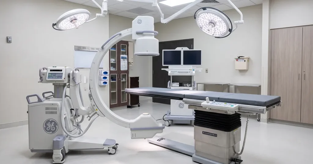
- GE OEC 9900 Elite Equipment
- All-Inclusive: Delivery, Setup & Retrieval
The Basic Idea Behind a C-Arm
The name comes from the shape: a C-shaped arm with an X-ray source mounted on one end and a detector on the other, positioned on opposite sides of the patient. X-rays pass through the body and get picked up on the far side, converted into a live image on a monitor. That part’s simple enough. What actually makes it work is four components doing their jobs in sequence, every time the pedal gets pressed.
The Four Components That Actually Make It Work
The X-Ray Generator
The generator is what produces the X-ray beam in the first place, and it’s rated in kilowatts and kilohertz, 15kW and 60kHz on the units Coast to Coast Radiology runs. Higher frequency generators produce a more consistent beam with less patient dose per image, which is part of why generator specs matter even though nobody outside biomedical engineering usually asks about them.
The Image Intensifier
On the detector side sits the image intensifier, a vacuum tube that converts the X-rays passing through the patient into a visible image. It comes in different sizes, 9-inch or 12-inch on the equipment Coast to Coast stocks, and the size determines the field of view: a smaller intensifier gives a tighter, more detailed picture, while a larger one captures more anatomy in a single shot. Choosing between them is a resolution-versus-coverage tradeoff, not a right-or-wrong answer, and it depends on the procedure.
The C-Arm Gantry
The gantry is the physical arm itself, and it’s built to rotate and swing around the patient without the patient moving, which is the entire point of the design. A technologist can angle the C into an AP, lateral, or oblique position mid-procedure, getting a different view of the same anatomy in seconds instead of repositioning the patient on the table.
The Monitor and Image Processing
The final piece is the display and the software processing what the intensifier captures. Newer units run articulating flat-panel monitors that show the live feed and often a reference image side by side, with digital processing cleaning up noise and enhancing edges before the image ever reaches the screen. This is also where stored images get saved for the patient record, rather than relying on film the way older fluoroscopy units once did.
How the Image Gets From the Patient to the Screen
Put those four pieces in order and the whole process looks like this: the generator fires an X-ray beam, the beam passes through the patient’s tissue and bone, the image intensifier on the other side catches what makes it through and converts that into a visible image, and the processing software cleans up and displays that image on the monitor in real time. All of that happens fast enough that the surgeon sees continuous motion, not a series of still frames, which is the difference between fluoroscopy and a single diagnostic X-ray.
Why the C Shape Matters for Positioning
A fixed X-ray unit takes one view from one angle. A C-arm’s whole value is that the gantry moves around a stationary patient instead of the other way around. During a spinal fusion or a complex orthopedic repair, a surgeon might need an AP view to confirm alignment, then swing to a lateral view to check hardware placement, all within the same case, without breaking sterile field or repositioning anyone on the table.
Picture a two-hour orthopedic case where hardware placement needs confirming from three different angles before closing. Without that rotating gantry, the team would be stopping to reposition the patient each time, adding time to a case where every extra minute under anesthesia matters. The C-arm just swings instead.
Built-In Radiation Dose Management
Modern C-arms don’t run a continuous X-ray beam. Most use pulsed fluoroscopy, firing short bursts instead of a steady stream, which cuts patient and staff dose significantly compared to older continuous units. Last-image-hold is another standard feature: the system freezes the most recent frame on screen so the surgeon can study it without the generator firing again. FDA’s equipment safety features for fluoroscopy cover this in more depth. Dose management deserves its own full breakdown given how much OR staff care about radiation safety day to day, and that’s a guide of its own.
What This Means When You're Renting
None of this matters much if the unit that shows up isn’t calibrated correctly, since a generator running slightly out of spec or an intensifier due for service shows up as inconsistent image quality mid-case, not as an obvious warning light. That’s why every C-arm Coast to Coast Radiology delivers gets calibrated and tested before it leaves the warehouse, not just plugged in and dropped off.
Frequently Asked Questions About How a C-Arm Works
What does the "C" in C-arm actually refer to?
It refers to the shape of the gantry, the physical arm that holds the X-ray source on one end and the image intensifier on the other, curved into a C so it can rotate around the patient.
Is a C-arm the same as a regular X-ray machine?
No. A standard X-ray machine takes a single still image. A C-arm produces a continuous, real-time image feed during a procedure, which is what makes it useful for guiding surgery as it happens rather than diagnosing after the fact.
What's the difference between the image intensifier size options?
A 9-inch intensifier gives a tighter field of view with more detail, while a 12-inch intensifier captures a wider area of anatomy in a single image. The right size depends on the procedure and the anatomy being imaged.
Does a C-arm expose the patient to more radiation than a standard X-ray?
A single fluoroscopy pulse is comparable to a standard X-ray exposure, but a procedure involves many pulses over time. Pulsed fluoroscopy and last-image-hold are both built in specifically to keep total dose as low as reasonably possible over the course of a case.
Who operates a C-arm during a procedure?
Typically a radiologic technologist positions and operates the unit while the surgeon directs what views are needed, though staffing setups vary by facility and procedure type.
How long does it take to set up a C-arm in a procedure room?
Setup typically runs 15 to 20 minutes once the unit arrives, covering positioning, power-up, and a quick function check before the first case.
Get a C-Arm That's Calibrated and Ready
Understanding how the machine works is one thing. Having a calibrated, ready-to-use unit show up on time is another. Coast to Coast Radiology delivers, sets up, and retrieves C-arm equipment across Florida, with the owners on the phone if anything comes up mid-rental. Call (941) 371-9659 or schedule online.